MB ChB 1994 Otago; FRACS 2005; ChM 2007 Sydney
Otago Medical School 1990-1994
MBChB 1994
FRACS Part I 1998
FRACS Part II 2002
FRACS 2004
Head and Neck Fellowship - Auckland Hospital ORL/Head and Neck 2004
Melanoma Fellowship - Sydney Melanoma Unit 2005
Head and Neck Fellowship - Sydney Head and Neck and Cancer Institute 2006
Master of Surgery in Surgical Oncology (University of Sydney) 2007
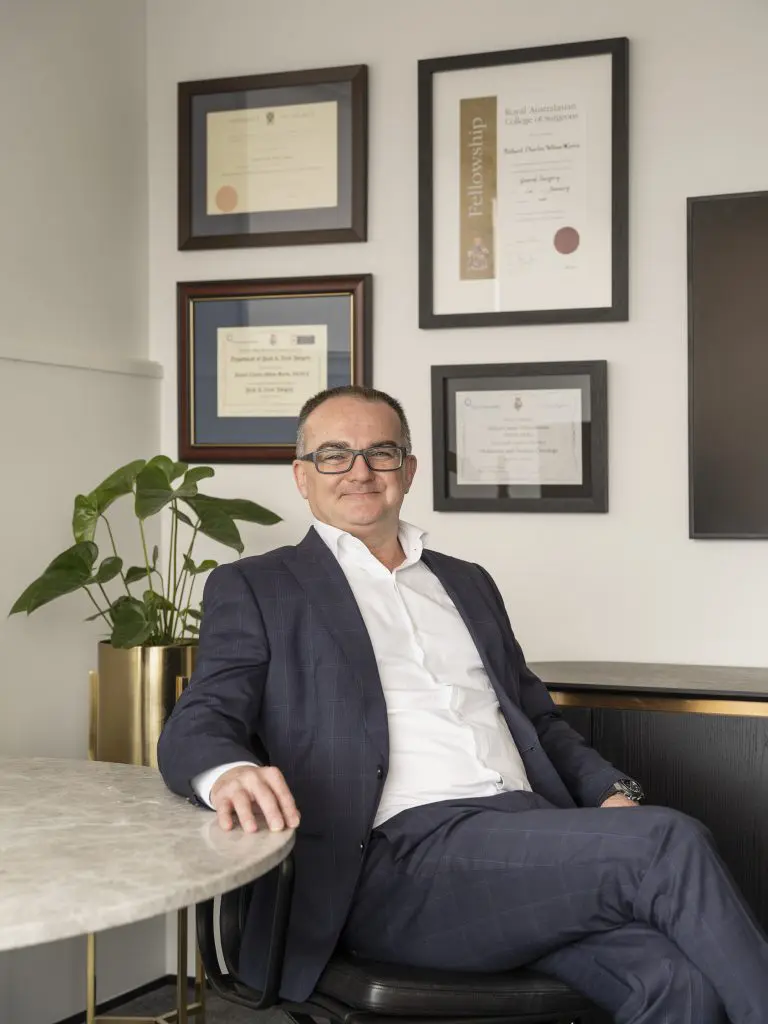
Richard Martin is a New Zealand trained general surgeon who spent two and a half years at the Sydney Cancer Center specialising in melanoma and head/neck surgery.
He is New Zealand's leading Melanoma Surgical Oncologist and key opinion maker.
Cutaneous Surgical Oncologist , Head/Neck/Endocrine and General Surgeon
Lorem ipsum dolor sit amet, consectetur adipiscing elit. Elementum eget vel, nunc nulla feugiat. Metus ut ultricies faucibus.
Book an AppointmentLorem ipsum dolor sit amet, consectetur adipiscing elit aliquet in
Lorem ipsum dolor sit amet, consectetur adipiscing elit aliquet in
Lorem ipsum dolor sit amet, consectetur adipiscing elit. Quam proin nibh cursus at sed sagittis amet, sed. Tristique id nibh lobortis nunc elementum. Tellus quam mauris aenean turpis vulputate sodales nullam lobortis. Vulputate tortor tincidun.
Lorem ipsum dolor sit amet, consectetur adipiscing elit. Quam proin nibh cursus at sed sagittis amet, sed.
Lorem ipsum dolor sit amet, consectetur adipiscing elit. Tristique libero viverra ultricies commodo.
Lorem ipsum dolor sit amet, consectetur adipiscing elit, sed do eiusmod tempor incididunt ut labore et dolore
Lorem ipsum dolor sit amet, consectetur adipiscing elit, sed do eiusmod tempor incididunt ut labore et dolore magna aliqua.
Lorem ipsum dolor sit amet, consectetur adipiscing elit. Quam proin nibh cursus at sed sagittis amet, sed.
Growths, lumps, tumours or masses on the head and neck can be benign (noncancerous) or cancerous and can form in the larynx, pharynx, thyroid gland, salivary gland, mouth, neck, face or skull.
• Neurological examination – assesses eye movements, balance, hearing, sensation, coordination etc
• MRI – magnetic resonance imaging uses magnetic fields and radio waves to give images of internal organs and body structures
• CT Scan – computer tomography combines x-rays with computer technology to give cross-sectional images of the body
• Biopsy – a sample of tissue is taken for examination under a microscope.
Lymph nodes in the neck often become swollen when the body is fighting an infection.
Noncancerous masses such as cysts are often removed surgically to prevent them from pressing on nerves and other structures in the head and neck.
Cancerous masses spread to surrounding tissues and may be:
• Primary – they arise in the head or neck. Mostly caused by tobacco or alcohol use•
Secondary – they have spread from a primary tumour in another part of the body.
Cancers may be treated by a combination of radiotherapy, chemotherapy and surgery.